|
|
Alignment
of innominate bones in physiatric practice considering
a reversible triad of pelvic obliquity, leg length difference, and scoliosis: a consecutive case-series study Jussi Timgren, MD, Physiatrist Abstract Context— Pelvic
obliquity, leg length difference, and
scoliosis present themselves as a reversible triad
among patients with musculoskeletal pain. Objectives—The purpose of this study
was to examine the prevalence of reversible pelvic obliquity, and to assess
the manifestations of asymmetry in skeletal posture. Setting—This study was set in the physiatric
practice by one practitioner. Patients—All
consecutive first-visit patients during one calendar year, totaling 563, seeking relief for diverse manifestations
of pain or discomfort were included in the present study. Interventions—When the practitioner
diagnosed pelvic obliquity, the patients were instructed to perform an alignment restoring self-correcting
muscle energy maneuver. Main Outcome Measure(s)—The practitioner examined
the patients in the standing, prone, and supine position and compared the
level of the iliac crests, inferior scapular angles, and anteroposterior
shoulder position. With the patients in the standing position, the level of
the iliac crests and scapular angles was determined either by using a
Palpation Meter® or manually. During the second visit, the maintenance of the
symmetry and degree of pain were evaluated. Results—Reversible pelvic
obliquity was found in 553 patients out
of 563 (98.2%), and all except one were able to
establish innominate alignment during the visit. The ilium was
anteriorly rotated in 45.1% of the patients, causing ipsilateral leg
lengthening and compensating scoliosis with a contra-lateral convexity. Among
the patients, 53.1% presented with upslip of the ilium associated with leg
shortening and scoliotic convexity, both ipsilateral. Of the 377 patients
coming to the follow-up visit, 82,2 % reported
significant or moderate improvement on their
functional ability and reduction of pain. The maintenance of the symmetry was significantly associated with the
alleviation of the symptoms. Conclusion—Reversible pelvic
obliquity is very common but mostly overlooked in patients having
musculoskeletal pain. It presents itself in two clearly different forms. The
restoration of innominate symmetry using the patients’ muscle energy is
practicable and does not require any manipulative skills. Introduction As early as in 1936, orthopedic surgeons Pitkin and
Pheasant described a malalignment syndrome of the pelvis, which they
called “Sacrarthrogenetic telalgia”. Since then, numerous studies
have evaluated sacroiliac joint dysfunction, scoliosis,
and leg length discrepancy as separate
factors. However, the possible interdependency of these three signs has received little attention in medical research.
Hjul et al. In 1980, DonTigny
proposed that anteriorly rotated dysfunction
of the sacroiliac joint is causing pelvic obliquity, a high iliac crest, an
apparent lengthening of the ipsilateral leg, and a laterally deviated lumbar
spine contribute to the etiology of the low back pain. Based on his
experience, he proposed that this condition could be corrected by
mobilization of the SIJ. A dysfunctional sacroiliac joint is widely recognized to be
a source of low back pain. The purpose of this
consecutive case series study was to examine the prevalence reversible pelvic
obliquity facilitated by the practitioner using the patients’ muscle energy and to assess the manifestations of
asymmetry in skeletal posture. Methods Experimental
design This study consists of a retrospective
analysis of all first visits to one physiatrist during the calendar year from January to December 2010.
The practitioner assessed the pelvic, scapular, and
shoulder position of all the patients. If pelvic obliquity was diagnosed, the patients were instructed
to perform an alignment restoring
self-correcting maneuver, after which the status of the pelvis and scapulae was reassessed. During a
follow-up visit, the practitioner repeated the previous assessments
requesting the patients to evaluate their pain and functional ability. Subjects The total number of
patients was 563 and 59 % of
them were female. The age distribution was between 7 and 89 years, the mean age being 46 years. Instruments In some of the patients, the difference in the level of the
iliac crests and inferior scapular angles was
measured using a Palpation Meter,
PALM® (Performance Attainment Associates, St. Pail, MN, USA), and in the rest of the patients, the manual assessment
was used. The Palpation Meter has been found to be a valid, reliable, and precise instrument for measuring the scapular position Procedures The practitioner noted
the medical history and examined the
patients in the standing, prone, and supine position. The level of the iliac crests,
inferior scapular angles, and anteroposterior shoulder position were compared. When the iliac crest was higher on one side, a
12-mm thick wooden plate lift was placed under both feet for lift. The leveling of the pelvis with a lift under one
foot but increased elevation with the same lift under the opposite foot further verified the existence of pelvic
obliquity. The practitioner repeated the assessment after the self-correcting muscle energy maneuver described by both DonTigny
The pelvic obliquity
was considered to be reversible when after the mobilization, the iliac
crests were in level and the same lift under both feet in turn equally
elevated both iliac crests. During the follow-up visit, the
patients were asked to evaluate their treatment
using a semi-quantitative scale from 3 to 1 as follows: 1) significant
improvement (patient estimated as having over
50% alleviation of symptoms), 2)
moderate improvement, and 3) no
response. The practitioner
performed Adam’s Forward Bend Test by patients by whom visible spinal
asymmetry remained after establishing the innominate equilibrium. Statistical analysis: Maintenance of symmetry was correlated with the treatment response by a 4-field matrix (improvement, no
improvement, maintenance of symmetry, or relapse of asymmetry). Statistical
analysis was performed using a -test. The present study focused on the innominate equilibrium and comorbidity-related
symptoms caused by intervertebral discs,
joints, nerves, or myofascial trigger points were not considered. Results The localization of pain
in individual patients varied from lower and upper back, neck, head, and the four extremities. The
practitioner interpreted the pain to be mainly of myofascial origin. The
duration of the presenting symptoms was definable in 506 of the 563 patients
(89,9%). The average length of symptoms was 4.7 years (median, 2.0
years). The pelvic
obliquity manifested itself in two
clearly different ways. The
practitioner defined them as anterior rotation and upslip of the ilium. Table
1 shows the observed differences between the two categories. The most noteworthy difference between the two is
that in the anterior rotation,
the 12-mm lift under the ipsilateral leg caused the iliac crest to elevate further, while in the upslip, the
placement of the lift caused the iliac crest to descend to the level of the
opposite side. Table 2 shows the mutual relationship between the two categories. Table1.
Anatomical differences between anterior rotation and upslip of the ilium
Table 2. The
number and mutual relation of anterior rotation and upslip
The change
from obliqueness to symmetry and vice versa was clear and without transitional forms. In both cases, establishing
the pelvic symmetry also caused restoration of equal leg length and straightening of
scoliosis. The asymmetry was
measured using the Palpation Meter in 130 patients (23.5%). The measured
height difference between the iliac crests
varied between 28 and 8 mm, with an average of 17 mm. The difference in height of the scapular lower angles varied between
22 and 0 mm, with an average of 13 mm. After re-establishing of the symmetry,
the difference between iliac crests was
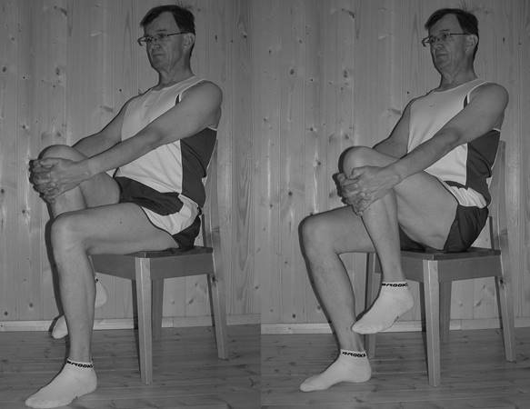
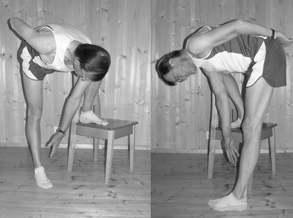
≤ 3mm. Both methods described in Figure 1 and 2 were equally effective
in re-establishing the symmetry. Seven patients with knee or hip prosthesis
used the latter method. In 4
cases, there was a concomitant idiopathic scoliosis with a positive Adams
Bend Test (e.g., unilateral elevation of thorax during spinal flexion). In
reversible pelvic asymmetry, the Adams test is negative. Idiopathic scoliosis
seldom causes rotatory component with protruding shoulder whereas that is the
rule in reversible scoliosis. By 28 patients
scoliosis and by 20 patients a leg length difference had been clinically
diagnosed in the past, up to 50 years earlier. Nine of the patients mentioned
above were given both diagnoses. By all of these patients, both scoliosis and
leg length turned out to be reversible. Follow-up Of all the 563 patients
377 (67,0%) attended the follow-up visit. The
average time between the first and the follow-up
visit was 44 days. One-hundred and eighty-one patients (48.0%) who attended
the follow-up visit reported a significant improvement in functional ability and reduction of pain, and
139 (36.9%) reported moderate improvement. The number of patients who
reported no improvement was 57 (15.1%). During the follow-up
examination by 354 (93.9%), pelvic symmetry had been maintained. Table 3. presents the maintenance of symmetry in correlation with
the treatment response by a 4-field
matrix (improvement, no improvement, maintenance of symmetry, or relapse of
asymmetry.) Treatment
response was strongly associated with the maintenance of symmetry ( = 32.7, df = 1, P-value < 0.001) Table 3. Correspondence of maintained symmetry and
significant or moderate improvement of condition reported by the patients
during their last follow-up visit.
Discussion The universality of the reversible innominate asymmetry is the most notable outcome of the present study. Analogous observations do exist. In our
previous study Schamberger Minor anatomical
asymmetries of the bony structures are very common in the normal population.
Ferreira et al. In the present study, no observable anatomical leg
length differences were found. It does not seem feasible
to prescribe shoe lifts to patients before a reversible pelvic asymmetry has been
ruled out. There were also patients in whom scoliosis had been diagnosed
years before in their school years, which now proved out to be of reversible
nature and others using prescribed shoe lifts with the assumption that the
leg length discrepancy is anatomical. The two major causes of pelvic obliquity anterior rotation and upslip
can be distinctly separated under two different conditions
although both share an anterior rotatory component of the ilium in the
standing position. Correspondingly, DonTigny The movement of the
sacroiliac joint is small and consists of a combination
of rotation and translation. The
rationale for the effectiveness of both
realigning maneuvers (Figures 1 and 2)
seems to be the fact that both share a
forced pull from the inferoposterior
region of the ilium through the gluteus maximus and hamstring muscles. The
attachment of the former is in the inferior
iliac spine and the latter in the ischial tuberosity. The maximal energy used first unlocks the location on the sacroiliac joint, from its maximum anterior rotation, and brings the
joint in its physiological middle position. The second maneuver causes an alternating stretch of the
same muscles. Both movements result in bringing the anteriorly rotated or up
slipped ilium back to the correct position. Based on
the present study findings, the sacroiliac joint
dysfunction is concurrent with the distorted position of the pelvis that can
easily be observed and measured. Whether this could contribute to the diagnosis of sacroiliac
joint dysfunction remains to be elucidated. This observational study did not
consider other co-existing factors of pelvic obliquity,
such as torsion of the sacrum and innominate inflare
or outflare. Conclusion Despite the high frequency of
reversible pelvic obliquity among patients having musculoskeletal pain,
pelvic obliquity remains mostly unrecognized. It presents itself in two
clearly identifiable but different forms.
The validity of the manual assessments can also be questioned. However, the described use of
the lift under both feet in an alternating sequence greatly enhances the
diagnostic accuracy. Many patients participating in this study had been seeking help repeatedly for years from diverse sources of
treatment in vain, which had led to frustration. However, the relief of
long-lasting pain after re-establishing pelvic
symmetry justifies considering it
primarily to be a consequence of pelvic obliquity. The
restoration of pelvic symmetry, leg length equality, and straightening of
scoliosis confirms their functional character. Instead of focusing primarily
on the dysfunctional sacroiliac joint as a potential cause of pain, the whole
picture, however, can first be attained when we consider the pelvis as a
functional unit affecting the spine and lower extremities. Acknowledgements: The author thanks Mikko Aronniemi, Ph.D for
statistical advice and an anonymous editor for revising the language. |
|
References
|
Pitkin H, Pheasant H.
Sacrarthrogenetic telalgia II. A Study of Sacral Mobility. Am J Bone Joint Surgery. April 1936;18(2):365-374. |
|
|
Juhl JH, Cremin TM, Russell G.
Prevalence of Frontal Plane Pelvic Postural Asymmetry—Part 1. The Journal of the
American Ostheopathic Association. October 2004;104(10):411-421. |
|
|
3. |
DonTigny. Anterior Dysfunction
of the Sacroiliac Joint as a Major Factor in the Etiology of Idiopathic Low
Back Pain. Physical Therapy. April 1990;70(4):250-262. |
|
4. |
Schamberger W. The
Malalignment Syndrome. London: Churchill Livingstone; 2002. |
|
Slipman C, Whyte W, Chow D, Chou
L, Lenrow D, Ellen M. Sacroiliac Joint Syndrome. Pain Physician. 2001; 4(2):143-152. |
|
|
Simopoulos T, Manchikanti L, Singh V, et al. A Systematic Evaluation of Prevalence and Diagnostic
Accuracy of Sacroiliac Joint Interventions. Pain Physician. May/June
2012;15(3):E305-E344. |
|
|
Behdad HR, Sharwin T, Hamilton
C, Danielle P. Diagnosis and Current Treatments for Sacroiliac Joint
Dysfunction: A Review. Current Physical Medicine and Rehabilitation Reports. Mar 2014;2(1):48-54. |
|
|
8. |
van der Wurff P, Meyne W,
Hagmeijer R. Clinical tests of the sacroiliac joint, A systematic
methodological review. Part 2: Validity. Manual Therapy. May
2000;5(2):89-96. |
|
Berthelot JM, Labat JJ, Le
Goff B, Gouin F, Maugars Y. Provocative sacroiliac joint maneuvers and
sacroiliac joint block are unreliable for diagnosing sacroiliac joint pain.
Joint
Bone Spine. Jan 2006;73(1):17-23. |
|
|
Rondeau W. The Accuracy of
the Palpation Meter (PALM) for Measuring Scapular Position in Overhead
Athletes, Dissertation. Chapel Hill: University of North Carolina; 2007. |
|
|
da Costa B, Armijo-Olivo S,
Gadotti I, Warren S, Reid D, Magee D. Reliability of scapular positioning
measurement procedure using the Palpation Meter (PALM). Physiotherapy. Mar 2010;96(1):59-67. |
|
|
Petrone M, Guinn J, Sutlive T,
Reddin, A, Flynn TW, Garber MP. The accuracy of the Palpation Meter (PALM)
for measuring pelvic crest height difference and leg length discrepancy. The Journal of
Orthopaedic and Sports Physical Therapy. Jun 2003;33(6):319-325. |
|
|
Hagins M, Brown M, Cook C, et
al. Intratester and Intertester Reliability of the Palpation Meter (PALM)
in Measuring Pelvic Position. The Journal of Manual & Manipulative Therapy. 1998;6(3):130-136. |
|
|
Azevedo D, Santos H, Carneiro
RL, Andrade GT. Reliability of sagittal pelvic position assessments in
standing, sitting and during hip flexion using palpation meter. Journal of Bodywork and
Movement Therapies. Apr 2014(2):210-214. |
|
|
15. |
DonTigny. A detailed and
critical biomechanical analysis of the sacroiliac joints and relevant
kinesiology: the implications for lumbopelvic function and dysfunction. In:
Vleeming A, Mooney V, Stoeckart R, eds. Movement, Stability &
Lumbopelvic Pain. 2nd ed. Edinburgh: Churchill Livingstone; 2007. |
|
Timgren J, Soinila S.
Reversible pelvic asymmetry: an overlooked syndrome manifesting as
scoliosis, apparent leg-length difference, and neurological symptoms. Journal of Manipulative
and Physiological Therapeutics. September 2006;29(7):561-565. |
|
|
17. |
|
|
18. |
Ferreira E, Duarte M,
Maldonado E, Barsanetti A, Marques A. Quantitative assessment of postural
alignment in young adults based on photographs of anterior, posterior, and
lateral views. Journal of Manipulative and Physiological Therapeutics. July/August
2011;34(6):371-380. |
|
Stovall B, Kumar S. Anatomical
Landmark Asymmetry Assessment in the Lumbar Spine and Pelvis: A Review of
Reliability. PM&R. January 2010;2(1):48-56. |
|
|
20. |
Brady RJ, Dean JB, Skinner TM,
Gross MT. Limb length inequality: clinical implications for assessment and
intervention. Journal of Orthopaedic & Sports Physical Therapy. May 2003;33(5):
221-234. |
|
21. |
Lee D. The Pelvic Girdle.
Second Edition ed. Edinburgh: Churchill Livingstone; 1999. |